

Understanding the science behind tooth extraction, bone preservation, and preparing for a dental implant in Phoenix.
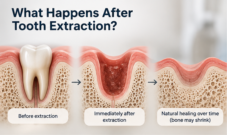
When a tooth needs to be removed, the goal isn't simply to extract it. It is important to plan for what happens to the surrounding bone and gum tissue afterward.
Following tooth extraction, the jawbone naturally begins to remodel and heal. Without the tooth's root, the surrounding bone will gradually lose height and width. These changes can affect your smile's appearance and make future dental implant placement more complicated.
At Innovative Dentistry Biltmore in Phoenix, Dr. Jennifer Ebner takes a biological, evidence-based approach to tooth extractions and surgical healing. This includes evaluating whether platelet-rich fibrin (PRF), bone grafting materials, barrier membranes, or a combination may help preserve bone at the extraction site for a future implant or dental bridge.
These procedures serve different purposes, and understanding those differences helps patients make informed decisions.
Why Preserving Bone After an Extraction Matters
Your natural teeth help maintain the bone supporting them. Once a tooth is removed, the surrounding bone begins to change as part of the healing process.
This is particularly important when a patient is considering a future dental implant.
An implant requires sufficient healthy bone for correct positioning and long-term support. If substantial bone loss occurs, the success of the implant can be compromised. Then additional and costly bone grafting procedures become necessary before implant placement.
A procedure called alveolar ridge preservation, also referred to as socket preservation or bone grafting, aims to limit the bone loss.
A dentist will use regenerative techniques, such as bone grafting, to help maintain the bone after a tooth extraction, instead of letting bone recession that comes with healing, occur.
The objective is not to prevent all bone loss, which isn’t possible, but to encourage favorable conditions for healing and bone maintenance.
What Is PRF, and How Does It Work?
Platelet-rich fibrin, or PRF, is a biological material prepared from a patient's own blood.
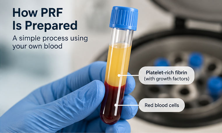
PRF uses naturally occurring components of the body's healing system. It uses the body’s own healing and grown factors right at the extraction site.
The process is straightforward.
A small sample of blood is collected, usually from the patient's arm.
The blood is placed into a centrifuge, which spins it at a controlled speed.
This separates the blood into components and allows a concentrated fibrin clot containing concentrated healing and grown factors to form.
The PRF can then be placed into the extraction socket, combined with grafting material, or prepared as a membrane.
PRF contains a fibrin network that release growth factors involved in normal tissue repair.
These biological components are shown to support early wound healing, soft tissue recovery, and avoid dry socket, a painful condition where the blood clot is lost.
Because PRF comes from the patient's own blood, it doesn’t contain and manufactured or donor materials. It alone does not stop the bone from remodeling and bone recession to occur.
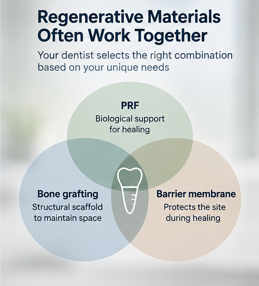
PRF provides biological support for healing. It does not function like a structural bone graft.
That distinction is important when bone preservation is needed for a future dental implant.
How Bone Grafting Materials Differ From PRF
Bone grafting materials provide a scaffold that supports new bone formation and helps maintain space as the extraction site heals.
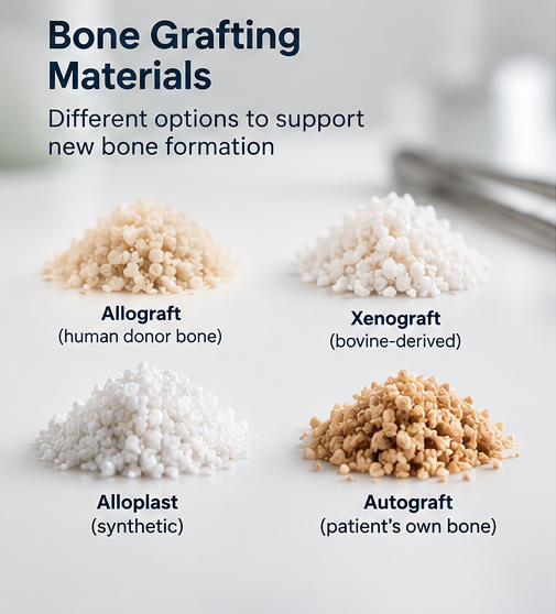
Several categories of bone grafts are used in modern dentistry.
Allografts are processed human donor bone materials. They provide a mineralized framework that can support natural bone remodeling.
Xenografts are derived from another species, commonly cow or pig bone, and undergo extensive processing. Some are particularly useful for maintaining ridge dimensions because they resorb slowly.
Alloplasts are synthetic bone substitutes, frequently made from calcium phosphate-based compounds.
Autografts come from the patient's own bone and may be appropriate for selected surgical defects but are usually painful to collect and also require healing.
Each material has different properties, including resorption rate, structural stability, and how it interacts with developing bone.
For patients interested in biological dentistry in Phoenix, material origin and bio-compatibility are reasonable considerations. Selecting a graft source should also consider the specific needs of the extraction site.
No single bone grafting material is ideal for every patient or every surgical situation.
What Are Barrier Membranes?
Barrier membranes perform a different function from either PRF or bone graft particles.
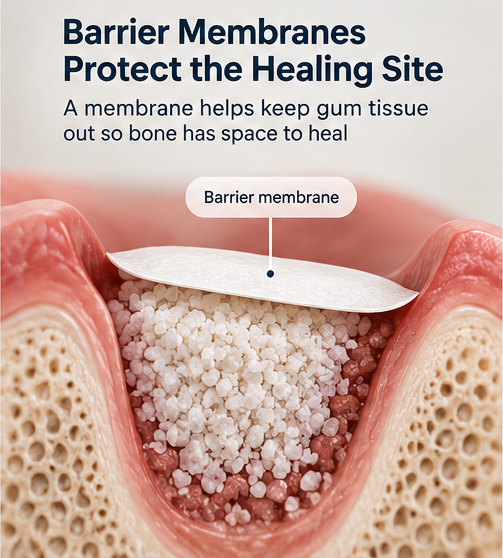
During healing, bone grafting particles can shift and gum tissue can grow into the extraction socket more quickly than bone develops. A barrier membrane helps separate these tissues, protecting the grafted area and maintaining space for bone healing.
This process is part of a technique known as guided bone regeneration.
Membranes may be made from teflon, metals, resorbable donated collagen or from part of the PRF clot. Some dissolve naturally during healing, while materials like teflon and metal require surgical removal.
The dentist combines a bone graft with a barrier membrane to help stabilize and protect the healing site.
PRF can also be formed into a membrane-like layer. However, a PRF membrane provides different barrier duration or structural stability than a collagen or teflon/ metal reinforced membrane.
The appropriate choice depends on the condition of the surrounding bone, gum tissue, and surgical defect.
What Does 2026 Research Show?
A systematic review and component network meta-analysis published September 18, 2026, in Frontiers in Oral Health examined 31 randomized controlled trials involving alveolar ridge preservation.
The researchers evaluated how different components, including grafting scaffolds, PRF, and barrier membranes, contributed to preserving jawbone dimensions after extraction.
These findings suggest that barrier membranes and allograft materials may make substantial contributions to preserving bone width, while PRF may provide an additional, smaller benefit.
These estimates were generated through statistical modeling. They aren’t guarantees or proof that one material is universally superior.
The authors also identified limitations, including inconsistent trial findings, limited comparisons, and low or very low certainty for several component-level estimates.
An additional 2026 meta-analysis comparing PRF directly with bone grafts found greater bone loss in PRF-only treatment groups. This supports the conclusion that PRF should not automatically replace structural grafting when preserving bone dimensions is a primary objective.
The findings indicate that PRF is a complement to bone grafting, and PRF alone isn’t sufficient to stop bone resorption while healing from an extraction.
How I Choose Regenerative Materials for Each Patient
At Innovative Dentistry Biltmore, regenerative planning begins with understanding the condition of the tooth and surrounding tissues.
Several factors influence my treatment recommendations.
The existing bone: A socket surrounded by intact bone walls requires a different approach than one affected by infection, periodontal disease, or significant bone loss.
The location of the tooth: Front teeth present important aesthetic considerations, while back teeth must tolerate substantial chewing forces and require different implant planning.
Future implant plans: If an implant is anticipated, maintaining adequate bone width and height influences the decision to recommend grafting.
The patient's healing considerations: Medical history, medications, smoking, periodontal health, and other individual factors will affect treatment choices.
Using three-dimensional cone-beam CT imaging before extractions helps evaluate bone anatomy and future implant planning.
In some situations, PRF alone may be recommended. In others, bone grafting with a protective membrane is more appropriate. A combination may offer advantages when additional biological support is needed.
Not every extraction requires grafting, and not every patient needs PRF.
A biological approach means using materials that support healing, limiting unnecessary procedures, and basing treatment on the patient's clinical needs rather than following a one-size-fits-all protocol.
Planning for Healthy Healing and Future Dental Implants
The best time to discuss bone preservation is before a tooth is extracted.
Early planning allows patients to understand whether socket preservation could reduce the likelihood of needing more extensive bone augmentation later.
Even with careful surgical techniques, natural remodeling and recession will occur. No regenerative material can guarantee complete bone preservation or successful future implant placement.
For patients seeking biological tooth extraction, bone grafting, or dental implants in Phoenix, I aim for the most appropriate procedures to get the results needed for the patients goals for their mouth.
Schedule a Biological Dentistry Consultation in Phoenix
If you have a tooth that needs extraction, or you're considering bone grafting or dental implants, understanding your options before treatment will make a meaningful difference.
At Innovative Dentistry Biltmore, Dr. Jennifer Ebner combines decades of clinical experience with modern imaging, biological techniques, and individualized surgical planning.
We welcome patients throughout Phoenix, Biltmore, Arcadia, Scottsdale, and the surrounding communities who want thoughtful, evidence-based dental care.
Schedule a consultation with Innovative Dentistry Biltmore to discuss your extraction, PRF, bone grafting, and dental implant options, and develop a treatment plan designed around your oral health and long-term goals.
Research references
1. Zhang J, Zhong J, Huang S, Zhang L. Component network meta-analysis of scaffold materials, platelet-rich fibrin and barrier membranes for alveolar ridge preservation. Frontiers in Oral Health. September 18, 2026. Read the study.
2. Platelet-rich fibrin versus bone grafts for alveolar ridge preservation: a systematic review and meta-analysis of randomized clinical trials. International Journal of Oral and Maxillofacial Surgery. August 2026. Read the study.
3. Reis NTdA, et al. Comparative efficacy of platelet-rich fibrin protocols for alveolar ridge preservation: a systematic review and network meta-analysis. BMC Oral Health. September 18, 2026. Read the study.